PC-SERS
By developing photonic crystal (PC) surfaces that incorporate metals and metal nanoparticles, the Nanosensors Group demonstrated PC-SERS as a means for increasing the electric field intensity exposed to metal surfaces, thereby increasing the detection sensitivity of SERS (1). More recently, we have been developing fabrication approaches for SERS nanoparticle surfaces that do not require photolithography, so the surfaces can be applied inexpensively over large surface areas and incorporated into single-use disposable laboratory items such as plastic tubing, microscope slides, and microplates.
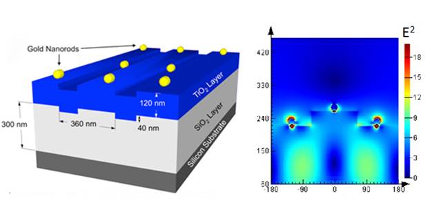
The amplification of the signals in SERS comes mainly through the electromagnetic interaction of light with certain metals (Ag, Au, Cu), which produces large amplifications of the laser field through excitations generally known as localized surface plasmon resonances.
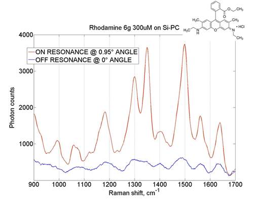
Photonic crystal surfaces coupled with metal nanoparticles provide a new mechanism for enhancing the electric field available for SERS. Our line-scanner microscope allows us to shine a laser at an angle on the photonic crystal surface in order to match its resonance mode.
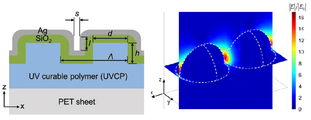
Nanodomes
This group developed a new SERS substrate consisting of a closely spaced metal nanodome array fabricated on flexible plastic film. We use a low-cost, large-area replica molding process to produce a two-dimensional periodic array of cylinders that is subsequently coated with SiO2 and gold (or silver) thin films to form dome-shaped structures with plasmonic properties.
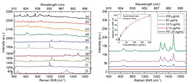
We have experimentally demonstrated that an in-line SERS sensor has a great potential for enhanced safety of current smart infusion systems through point-of-care detection and real-time monitoring of intravenous drug delivery within biomedical tubing.
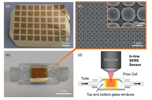
The ability of SERS sensors to quantitatively identify drug compounds, kinetically monitor changes in drug concentration, and simultaneously detect a combination of two drugs has been demonstrated:
References
- (*W) “Photonic crystals with SiO2-Ag “post-cap” nanostructure coatings for surface enhanced Raman spectroscopy,” S.-M. Kim, W. Zhang, and B.T. Cunningham, Applied Physics Letters, Vol. 93, p. 143112, DOI: 10.1063/1.2998695, Published Online October 9, 2008.
- “Surface-enhanced Raman nano domes,” C.J. Choi, A. Xu, H.-Y. Wu, L. Liu, and B.T. Cunningham, Nanotechnology, Vol 21, p. 415301 (2010) DOI: 10.1088/0957-4484/21/41/415301.
- “Plasmonic coupling of SiO2-Ag ‘post-cap’ nanostructures and silver film for surface enhanced Raman scattering,” H.-Y. Wu and B.T. Cunningham, Applied Physics Letters, Vol. 98, Issue 15, p. 153103, 2011 (Featured on Cover).
- “Quick detection of contaminants leaching from polypropylene centrifuge tube with surface-enhanced Raman spectroscopy and ultraviolet absorption spectroscopy,” Z. Xu, C.J. Choi, B.T. Cunningham, and G.L. Liu, Journal of Raman Spectroscopy, published online April 19, 2011, DOI:10.1002/jrs.2950.
- “Biochemical Sensor Tubing for Point-of-Care Monitoring of Intravenous Drugs and Metabolites,” C.J. Choi, J. Weyhenmayer, H.-Y. Wu, and B.T. Cunningham, Lab on a Chip, Vol. 12, No. 3, p. 574-581, 2012.
- “Nanoreplicated positive and inverted submicron polymer pyramids array for Surface-Enhanced Raman Spectroscopy,”,” Z. Xu, H.-Y. Wu, S.U. Ali, J. Jiang, B.T. Cunningham, and G.L. Liu, Journal of Nanophotonics, Vol. 5, p. 053526, 2011. (doi: 10.1117/1.3663259)
- “Plasmonic nanogap-enhanced Raman scattering using a resonant nano-dome array,” H.-Y. Wu, C. J. Choi and B.T. Cunningham, Small, Vol. 8, No. 18, p. 2769, 2012 (Featured on Cover).
- “Point-of-care detection and real-time monitoring of intravenously delivered drugs via tubing with an integrated SERS sensor,” H.-Y. Wu and B.T. Cunningham, Nanoscale, Vol. 6, p. 5162, 2014 (DOI: 10.1039/C4NR00027G)