AirPods-sized fluorescence analytical device holds the promise for timely home molecular testing

BY CLAUDIA LUTZ

July 15, 2026

Pictured left to right: Skye Shepherd, Han Keun Lee, Amanda Bacon, My Thi Tra Nguyen, and Brian Cunningham

Pictured left to right: Skye Shepherd, Han Keun Lee, Amanda Bacon, My Thi Tra Nguyen, and Brian Cunningham / Isaac Mitchell

Advances in medical technology have improved our health in part by bringing key aspects of care, once difficult to access, into the home. Tracking symptoms and even screening for certain types of illness outside of a laboratory or clinical setting puts more control into the hands of patients. New research from the University of Illinois is helping to provide a practical answer to the question, what does a holistic system need to look like in order to work outside the laboratory?

Research led by Han Keun Lee in the laboratory of electrical and computer engineering professor Brian Cunningham (CGD leader), in collaboration with bioengineering professor Xing Wang (CGD), was published in the IEEE Sensors Journal. Lee, Cunningham, Wang, and their coauthors shared the design and validation of a device that offers the capability to read and compare results from a variety of sensitive tests for pathogens or cancer-associated biomarkers, all within a housing similar in size and shape to an AirPods case. The study was supported by the National Institutes of Health and the U.S. Department of Veterans Affairs.

“I have an ambition that we can bring cancer detection to the home. Currently, detecting cancer often requires going to a hospital and having blood drawn,” requirements that can become barriers to accessing timely care, Lee said. “My job here is to be able to bring those tests out to the world so people can start utilizing them and have better access to the state-of-the-art technologies.”

Han Keun Lee displays an AirPods case (left) along with the similarly-sized VPod (rear) and VPodDuo (right) testing devices.

Many home tests work similarly to pregnancy tests or infectious diseases tests such as COVID-19 antigen tests; labeled molecules generate a visible line when a substance of interest is present in the sample, producing a simple positive or negative result. This type of test is relatively affordable and easy to use, but it can have limited sensitivity and often provides only qualitative or semi-quantitative results. A new version of the test strip must also be developed for each new pathogen or biological molecule of interest.

Another type of test, commonly used in the laboratory, can be much more accurate and quantitative because it relies on the measurement of fluorescent dye whose signal can be amplified. But many existing instruments used to read fluorescent signals are not practical for home or point-of-care settings; they are large, expensive, and often require training to use correctly.

“There are many different ways of quantifying fluorescent assays. One example is using a camera to capture the whole reaction area. . . but this requires sophisticated instrumentation,” Lee said. “That was deviating from our point of view where we wanted something that’s very portable, small and inexpensive. We decided to stick with a photodetector,” a simple component that detects the intensity of light without capturing spatial information.

Using a simple light detector has its own drawbacks; a single detector does not allow a clear comparison between the sample that comes from the individual being tested and a control sample that provides a baseline negative result. Lee and his colleagues had previously designed a compact, user-friendly device they called the VPod that was able to detect fluorescence signals, but could measure only one sample at a time. Their present publication showcases their improved device, the VPodDuo, which supports a paired test-and-control workflow: this means that it is able to measure the two samples simultaneously.

VPodDuo, like the VPod before it, is also suited to point-of-care settings because of its compatibility with different molecular-assay formats. The device measures green-emitting fluorescence signals generated by several types of detection chemistry, allowing the same reader to be used with tests for different targets. The research team demonstrated that VPodDuo could accurately detect and measure quantities of genetic material from Zika virus, HIV, and methicillin-susceptible Staphylococcus aureus bacteria, as well as human genetic markers indicating the possible presence of cancer cells.

“There is a famous saying that medicine is blind without diagnostics,” Lee said. “The purpose of testing at the point-of-care is not necessarily to give a definitive diagnosis, but rather to allow more frequent testing so that someone has a better chance of receiving timely treatment.”

The VPodDuo’s versatility and accuracy make it a valuable piece of scientific equipment. To be practical for point-of-care applications, it also needed to be portable and easy to use. Lee and his coauthors designed the device to connect wirelessly to a mobile device, designed a software application with an intuitive user interface to aid with operation and result intepretation, and built in safeguards to protect against accidental misuse. All of these features are directed toward their overall goal of creating a point-of-care testing workflow that can be used outside of centralized laboratories.

“It’s not just about the individual test for point-of-care use, it’s about the entire system,” Lee said. “We wanted to address this from a system-level engineering perspective for fluorescent molecular testing. That’s where I wanted to go, so that I can help bring all these great technologies out into the world to provide real benefit.”

Paradigm Shift in Oncology: CCIL Lab Pioneers Multi-Institutional Publication On Critical Emergence of Point-of-Care Diagnostic Technology

Jul 13, 2026 | Cancer Center News

Who doesn’t want cancer testing results at less cost, with greater speed and accuracy? Interdisciplinary innovators like Cancer Center at Illinois (CCIL) Program Leader Brian Cunningham are among the pioneers of the emerging field of point-of-care (POC) diagnostic technology aiming to make this dream a reality.

To present the case for the transformative shift in oncology underway thanks to POC molecular diagnostic technology, Cunningham initiated a multi-institutional, interdisciplinary collaboration. The team published a critical review paper arguing POC technology stands to dramatically change the future of cancer detection, therapy selection, therapy effectiveness monitoring, and personalized medicine.

For the paper, Cunningham organized clinical and engineering collaborators from the CCIL, University of Illinois Chicago, Huntsman Cancer Institute, Mayo Clinic, and Stanford School of Medicine. The team’s critical review paper is published in the Royal Society of Chemistry’s Lab on a Chip journal, and the issue’s cover photo features liquid biopsy POC technology developed in Cunningham’s lab that can produce oral cancer results in less than 15 minutes.

Leveraging case studies and clinical scenarios for oral squamous cell carcinoma, hepatocellular carcinoma, prostate cancer, and non-small cell lung cancer, the team explains both the challenges and opportunities of POC diagnostic technology. Challenges include funding and regulatory approval, sample collection methodology variations, and technology manufacturing and commercialization. But these challenges are part and parcel of any technology revolution.

Cunningham suggests this paradigm shift in the field of molecular diagnostics holds echoes of the transition from film to digital photography. In the era of film photography, the reader may recall, a photographer would have to ship off film to a central processing facility at higher cost and with long wait times. But with the advent of digital photography, a photographer can now view her captures rapidly and at less cost and often with much greater clarity. Similarly, the dependency upon centralized laboratory testing is a major obstacle in cancer detection, therapy, and treatment monitoring.

The Lab on a Chip journal featured point-of-care diagnostic technology developed in Cancer Center at Illinois researcher Brian Cunningham’s lab.

“Today, many molecular diagnostic tests still rely on centralized labs and delayed turnaround times, whereas the vision outlined in this work is a future where clinically actionable molecular info can be obtained much closer to the POC, enabling faster treatment and management decisions,” said Seemesh Bhaskar, first author of the team’s paper.

Cunningham’s lab is contributing to this POC revolution in numerous ways, including:

  • Inventing, developing, and applying new biosensing platform technologies that have the capability for “digital precision” detection and counting of individual molecules and viruses.
  • Inventing novel biochemistry methods that work with the biosensing technologies to make diagnostic test workflows that are simple and fast.
  • Collaborating closely with clinicians who have specific but broadly important use cases for us to demonstrate in fields like cancer therapy selection, maternal health, nutrition, and veterinary diagnostics.

“The burden of cancer urges us to develop swift, precise, and accessible diagnostic responses that can overcome the limitations of diagnostic tests performed in centralized laboratory settings,” said Cunningham. “Point-of-care molecular diagnostics are emerging as transformative tools that enable decentralized testing across hospitals, outpatient clinics, oncology practices, and dental practices, thereby accelerating clinical decision-making to improve patient outcomes.”

Point-of-care cancer diagnostics holds promise for patients facing a variety of obstacles. Consider patients who experience considerable anxiety while waiting for test results to arrive, or patients who fail to make follow-up appointments for sample collection. Consider the barriers for people living in rural areas. Consider the lack of adequate health insurance or a fear of a cancer diagnosis that may lead many patients to avoid taking extra steps for diagnostic tests.

“Overall, there are many societal, personal, and financial barriers that lead to healthcare disparities,” reported Cunningham. “These issues could be partially addressed through the availability of low cost, rapid, noninvasive, and actionable molecular diagnostic tests that can be performed and interpreted during a visit to a physician’s office.”

By analyzing the global funding landscape, intellectual property demands, and regulatory frameworks that influence the transition of emerging technologies from academic research to commercial markets, in addition to the clinical and research lab case studies, Cunningham’s review not only summarizes the current state of the field but also provides a roadmap for translating POC cancer diagnostics into routine clinical practice. Such a review highlights the critical role of the basic science research and innovation emerging from the unique, interdisciplinary, cancer-engineering driven ecoystem at the Cancer Center at Illinois.

“POC molecular diagnostics for cancer is not a big part of healthcare yet,” concluded Cunningham. “The point of our paper is that there are big clinical needs and important scenarios where physicians want POC technology for specific purposes, and that there is technology developing that can address and remedy those needs.”

Cunningham (right) and postdoctoral researcher Seemesh Bhaskar analyze results from a laboratory experiment.

Editor’s notes:

The research team acknowledges the following funding sources. Seemesh Bhaskar acknowledges funding from the Carl R. Woese Institute for Genomic Biology (IGB). Ugur Aygun acknowledges funding from the European Union’s Horizon Europe research and innovation programme under the Marie Skłodowska-Curie grant agreement No. 101066038. Fatma Uysal Ciloglu is supported by the Scientific and Technological Research Council of Türkiye during her post-doctoral research.

Brian Cunningham is the Intel Alumni Endowed Chair in the Department of Electrical and Computer Engineering, a Program Leader at the Cancer Center at Illinois, an affiliate of the Department of Chemistry, the Department of Bioengineering, and the Holonyak Micro and Nanotechnology Laboratory, and a Working Group Leader for the CCIL and IGB’s collaboration at the Center for Genomic Diagnostics.

He can be reached at bcunning@illinois.edu.

The article “Point of care molecular cancer diagnostics” was published in the Royal Society of Chemistry’s Lab on a Chip journal and is available here.

DOI: doi.org/10.1039/d5lc01014d

This story was written by Jonathan King, CCIL Communications Coordinator.

The Power of Photonics

WRITTEN BY CASSANDRA SMITH – March 2026

Timing is critical in diagnosing diseases such as cancer. Researchers within The Grainger College of Engineering at the University of Illinois Urbana-Champaign used a historically underappreciated tiny powerhouse to detect diseases sooner. 

“I’m using all my knowledge for cancer diagnostics. That’s a shared mission between me and Professor Cunningham. ”

Seemesh Bhaskar

Seemesh Bhaskar believes cancer detection should happen years before a diagnosis ever appears in a medical chart. 

The postdoctoral researcher in Professor Brian Cunningham’s Nanosensors Group is helping develop technology that could detect signs of cancer five to eight years earlier than traditional diagnostic tools by identifying molecular signals long before symptoms emerge. 

Bhaskar is using his multidisciplinary academic background in physics, environmental diagnostics, photonics, chemistry and nanotechnology to pursue that goal.

“I’m using all my knowledge for cancer diagnostics. That’s a shared mission between me and Professor Cunningham,” Bhaskar said.  

THE RESEARCH

Their work recently appeared in Chemical Reviews — the highest-impact-factor journal in chemistry, published by the American Chemical Society. The paper represents more than two years of research, including extensive reviews of discoveries spanning from 1900 to 1980, to better understand why there is often a large gap between cancer’s initial development and its formal diagnosis. 

When it comes to disease, the timing of a diagnosis is critical. Through a combination of photonics and nanotechnology, their research could enable much earlier detection: five to eight years sooner than traditional instruments, according to Bhaskar. Their tiny technology packs a big punch.

Side-by-side photos of Brian Cunningham and Seemesh Bhaskar
Professor Brian Cunningham and Seemesh Bhaskar

“Everything boils down to DNA and RNA,” Bhaskar said. “They must get flicked out and do something wrong. And that will lead to cancer.” 

While the reason for a cell’s mutation remains under active study, the Illinois researchers can detect signals that may predict a cell’s mutation years later. 

“We wanted to see if we could trace it back to a point where something is happening in the cell, and can we do the real detection there?” Bhaskar said. “That is where the power of photonics comes.” 

Indeed, there is power in photonics, and it works on a microlevel. 

“We have microorganisms, viruses and bacteria in our systems that attack the body from external sources,” Bhaskar said. “They are very small. Molecules are very tiny. So we cannot see the molecules or bacteria because they are so small. The resolution is not high enough. So we need something that can talk to them.” 

Enter nanomaterials. 

Nanotechnology is small enough to interact with microRNA. Light and nanomaterials can interact with one another, and the nanomaterials can also interact with biological systems. 

“Photonics has this very strong power where we can indirectly talk to the molecules and see how they are creating problems in terms of how the disease is spreading,” Bhaskar said. 

Once researchers identify the problem area, health professionals can detect the disease and pursue treatment. 


SHEDDING LIGHT ON WHAT WAS IGNORED

What took so long to shed light on this powerful detection method? Bhaskar said researchers largely ignored it.

“Earth is a giant magnetic field. Light is called electromagnetic radiation,” said Bhaskar. “For so many decades, we only talked about electric flux. We totally ignored the magnetic flux of the electromagnetic radiation that is traveling. Simply because the magnetic flux cannot be accessed, there were not many accessible tools.”

Bhaskar and his team created simulations using nano-assemblies made in the laboratory that could tap into that magnetic flux potential. 

“We engineered the photonic substrates and the nano-assemblies and made them talk to each other and did the detection of microRNA,” Bhaskar said. “So, if a patient is going to get cancer five years down the line, their body is going to have a few microRNAs or DNA that are actually creating the cancer five years down the line.”

A LAB BUILT ON MENTORSHIP

Seemesh Bhaskar talks with professor Brian Cunningham during poster session
Seemesh Bhaskar talks with professor Brian Cunningham during poster session.

Publishing  in Chemical Reviews-the first time Bhaskar and Cunningham have appeared in the journal-is a source of pride for both. 

Bhaskar said the paper was successful in part because of the laboratory environment fostered by Cunningham. 

“He treats everyone like faculty,” Bhaskar said. 

Bhaskar said Cunningham takes a holistic approach to mentoring researchers. 

“Working with him is gripping,” Bhaskar said. “Once you talk to him about what you want to do, he will go back to the same point of how it is beneficial to the society.” 

Cunningham supports researchers personally by asking during team meetings about something positive that happened in their lives that week and showing genuine interest in their well-being. 

For Bhaskar, the goal of the research ultimately comes back to people. 

“If we can detect the signals earlier, we give doctors more time,” he said. “More time means more options for treatment and a better chance for patients.” 


Grainger Affiliations

Professor Brian T. Cunningham is an Intel Alumni Endowed Chair Professor in the Department of Electrical & Computer Engineering; Program Leader for the Cancer Center at Illinois in the Office of the Vice Chancellor for Research and Innovation; Professor in the Department of Bioengineering; Professor within the Holonyak Micro & Nanotechnology Lab; Professor of Biomedical and Translational Sciences; Professor in the Department of Nutritional Sciences;Affiliate in the Department of Chemistry;Professor in the Beckman Institute for Advanced Science and Technology;Affiliate Professor in the Carl R. Woese Institute for Genomic Biology.

Chemical Reviews: Photonic Crystal Guided Resonance in Healthcare

A century of photonics research shapes the future of healthcare diagnostics

BY KATIE BRADY

October 23, 2025

The authors highlighted their collaborative team effort by sporting their Illini Orange and Blue. From left to right: Hankeun Lee, Joseph Tibbs, Amanda Bacon, Takhmina Ayupova, Leyang Liu, Anqi Tan, Wang-Chien Chen, Saurabh Umrao, Seemesh Bhaskar, Brian Cunningham, Xing Wang / Isaac Mitchell

The authors highlighted their collaborative team effort by sporting their Illini Orange and Blue. From left to right: Hankeun Lee, Joseph Tibbs, Amanda Bacon, Takhmina Ayupova, Leyang Liu, Anqi Tan, Wang-Chien Chen, Saurabh Umrao, Seemesh Bhaskar, Brian Cunningham, Xing Wang / Isaac Mitchell

Share

Diagnostic testing is essential in modern healthcare settings. By sensing biological molecules called biomarkers, clinicians can gain insights into what is happening in the body, invisible to the eye. Recently, a collaborative team from the Carl R. Woese Institute for Genomic Biology published a comprehensive review on photonic crystal grating resonance, or PCGR, a promising technology for improving diagnostic biosensors and early disease detection. 

The review article, published in the journal Chemical Reviews, provides a comprehensive overview of PCGR-based biosensors and their widespread applications—from pathogen detection and wearable diagnostics to environmental monitoring and toxin biosensing. It also dives into the past century of research that built the foundation for today’s technology and future advancements.

“It cites landmark works from 1907, 1947, 1956, and many more, connecting them to current breakthroughs in microRNA detection, lipid biosensing, circulating tumor DNA diagnostics, and beyond,” said Seemesh Bhaskar, first author of the paper and an IGB Postdoctoral Fellow in Professor of Electrical and Computer Engineering Brian Cunningham’s (CGD leader) research group. “To understand where we are going, it is essential to know where we have been. This review stands as a reminder that scientific foresight begins with historical awareness.” 

The first grating photonic substrate was reported in 1902. Since then, these advanced nanomaterials have become much more sophisticated, with researchers designing and fabricating specialized substrates for different applications. Photonic grating crystals are particularly beneficial for diagnostic biosensing due to their ability to manipulate how light is absorbed and reflected on the nanoscale. This helps to amplify the biosensor’s fluorescence signal and uncover ultra-low levels of molecular biomarkers such as proteins, viruses, or nucleic acids.

“One of the real strengths of this review is not just its comprehensive content, figures, and references, but also its carefully curated tables,” said Saurabh Umrao, a postdoctoral researcher in Associate Professor of Bioengineering Xing Wang’s lab. The first table of the review, for example, outlines advancements in the field over the past 20 years. Drawing on insights from over 100 studies, it provides a comprehensive list of diagnostic technologies that use one-dimensional photonic crystals. 

“In that sense, the review feels almost like an encyclopedia for the field, making it a valuable resource for both experts and newcomers,” Umrao said.

The field of diagnostics is highly broad and interdisciplinary, requiring knowledge from many areas including biology, chemistry, biomedical engineering, and materials science. A strength of this review is its holistic perspective that touches on each of these different areas, with an emphasis on not only sharing relevant information, but also providing critical analysis by experts.

When beginning the process of drafting the review, Bhaskar and Cunningham quickly realized that it was building into a much larger project than initially planned. This led them to recruit more people, and what was initially a four-person project, grew into a large team effort that played to everyone’s strengths.

Bhaskar said, “We thought the best approach was to incorporate people who have expertise in that domain. Anybody who looks at it will get a comprehensive picture, and that was only possible by having multiple people’s experience.” Bhaskar, in particular, worked on the technical background of photonics and plasmonics and personally took an interest in historical perspectives, while Cunningham used his extensive knowledge for higher level analysis like addressing current challenges and future considerations for the field. 

With background at the interface of biology and engineering, Umrao found it rewarding to write the diagnostics sections and explore emerging photonics-based technologies. Other members of the team contributed sections focused on substrate engineering or added chapters on machine learning.  

“This collaborative approach is precisely what makes the review both comprehensive and authoritative, and I’m confident it will resonate with the community,” said Leyang Liu, a graduate student in Cunningham’s lab. Liu wrote a chapter on hybrid structures, focusing on plasmonic films, Bragg mirrors, and dielectric gratings. “It’s been a pleasure to contribute to this review, and the process has been energizing and genuinely enriching.” 

As modern research has become increasingly complex and driven by discipline-specific work, the team hopes the review article can also inspire future interdisciplinary collaboration to develop new diagnostics. “Places like IGB are the best. You are put in a place where you can talk to interdisciplinary researchers. When this becomes part of life for scientists, many new ideas can come up,” Bhaskar said.

Another key aspect for advancing future PCGR-based diagnostic technologies is to addressing current pitfalls. Bhaskar and his colleagues did this by discussing topics like biosensor fabrication limitations and regulatory gaps. By identifying these issues, it can help to spark conversations that eventually lead to creative and innovative solutions.

Bhaskar said, “With a vibrant team of co-authors and collaborators, and with the support of our research centers and departments, we are excited about the continued journey of PCGR technologies and their potential to transform health diagnostics for all.”

The publication, “Photonic Crystal Grating Resonance and Interfaces for Health Diagnostic Technologies” can be found at https://doi.org/10.1021/acs.chemrev.4c00653 and was supported by the National Institutes of Health and National Science Foundation.

Manipulation of light at the nanoscale helps advance biosensing

BY KATIE BRADY

July 1, 2025

(from left) Professor Brian Cunningham, Amanda Bacon , Joseph Tibbs , Leyang Liu, Seemesh Bhaskar, Weinan Liu  / Isaac Mitchell

(from left) Professor Brian Cunningham, Amanda Bacon , Joseph Tibbs , Leyang Liu, Seemesh Bhaskar, Weinan Liu  / Isaac Mitchell

Traditional medical tests often require clinical samples to be sent off-site for analysis in a time-intensive and expensive process. Point-of-care diagnostics are instead low cost, easy-to-use, and rapid tests performed at the site of patient care. Recently, researchers at the Carl R. Woese Institute for Genomic Biology reported new and optimized techniques to develop better biosensors for the early detection of disease biomarkers.

People have long been fascinated with iridescence of peacock feathers, appearing to change color as light hits them from different angles. With no pigments present in the feathers, these colors are a result of light interactions with nanoscopic structures, called photonic crystals, patterned across the surface of the feathers. 

Inspired by biology, scientists have harnessed the power of these photonic crystals for biosensing technologies due to their ability to manipulate how light is absorbed and reflected. Because their properties are a result of their nanostructure, photonic crystals can be precisely engineered for different purposes.

The Nanosensors Group at the University of Illinois Urbana-Champaign, led by Professor of Electrical and Computer Engineering Brian Cunningham (CGD leader), previously developed photonic crystal-based biosensors that amplify the fluorescence using gold nanoparticles, which act as tags for sensing various molecular biomarkers. But while this innovative technology enables low-level detection of biomarker molecules, it still has room for further improvement.

“Traditionally, metal nanoparticles, especially gold, offer the potential for fluorescence enhancement, but suffer from a fundamental flaw at close range,” said Seemesh Bhaskar, an IGB fellow in the CGD research theme and lead author of the study. “These nanoparticles can quench— or decrease—the very fluorescence signals they aim to amplify. This creates a dead zone of detection, limiting the sensitivity of biosensors.”

In a paper published in MRS Bulletin, the research team aimed to overcome this limitation by introducing a new class of cryosoret nanoassemblies; these organized structures comprised of gold nanoparticle subunits are formed via rapid cryogenic freezing. 

“Self-assembly is a fundamental principle of nature, whether it’s the formation of planetary systems in cosmology or the precise organization of nucleotides in DNA,” Bhaskar said. “What individual nanoparticles cannot accomplish alone becomes possible through their collective organization. At its core, it’s about engineering optical behavior—both structurally and functionally—through deliberate design.”

By integrating these cryosoret nanoassemblies with specially designed photonic crystals, the fluorescence demonstrated a 200-fold signal enhancement compared to the photonic crystal alone. This showed that fluorescence quenching was effectively minimized, making this technology a promising avenue for detecting low concentrations of biomarkers.

Building upon this work, the team next sought to introduce magnetic tunability into the nanoassemblies, with the long-term goal of developing intelligent, responsive biosensors.

Light is a specific frequency range of electromagnetic radiation; other examples include radio waves, microwaves, and X-rays. Electromagnetic radiation travels through space in the form of waves, and as its name suggests, consists of both electrical and magnetic components. While many biosensing systems take advantage of the electrical component of light, the magnetic component is largely overlooked.

In a study reported in the journal APL Materials, Bhaskar and his colleagues designed magneto-plasmonic cryosoret nanoassemblies. They integrated these nanoassemblies onto a photonic crystal interface and found that it successfully harnessed both the electric and magnetic components of light. They tested their platform using a common fluorophore, which resulted in ultra-sensitive detection in the attomolar range, while still minimizing fluorescence quenching. 

Overall, this dual-mode interaction allows for enhanced control over light-matter interactions at the nanoscale, offering a new method to design highly sensitive and tunable biosensing platforms.

“This work represents a hybrid optical platform where photons are not merely emitted—they are orchestrated,” Cunningham said. “This convergence of photonic-plasmonic simulations, advanced nanofabrication, and chemical engineering principles has far-reaching implications, particularly in the realm of medical diagnostics.”

Moving forward, the researchers plan to continue optimizing the cryosoret nanoassemblies to target specific biomarkers, like microRNAs, circulating tumor DNA, and viral particles, for early detection of cancer and infectious disease. They hope that with further improvement, point-of-care technologies can meet the pressing need for sensitive, accessible, and deployable biosensing systems.

The publication in MRS Bulletin “Photonic-crystal-enhanced fluorescence: Template-free gold cryosoret nanoassembly steering, dequenching, and augmenting the quenched emission from radiating dipoles” can be found at https://doi.org/10.1557/s43577-024-00850-2

The publication in APL Materials “Photonic crystal band edge coupled enhanced fluorescence from magneto-plasmonic cryosoret nano-assemblies for ultra-sensitive detection” can be found at https://doi.org/10.1063/5.0251312 

The work was supported by the National Institutes of Health, National Science Foundation, and Cancer Center at Illinois.

Machine learning method helps bring diagnostic testing out of the lab

BY KATIE BRADY

June 16, 2025

Graduate student in electrical and computer engineering Han Lee (left) and Professor Brian Cunningham (right)

Graduate student in electrical and computer engineering Han Lee (left) and Professor Brian Cunningham (right) / Julia Pollack

Share

What if people could detect cancer and other diseases with the same speed and ease of a pregnancy test or blood glucose meter? Researchers at the Carl R. Woese Institute for Genomic Biology are a step closer to realizing this goal by integrating machine learning-based analysis into point-of-care biosensing technologies. 

The new method, dubbed LOCA-PRAM, was reported in the journal Biosensors and Bioelectronics and improves the accessibility of biomarker detection by eliminating the need for technical experts to perform the image analysis.

Traditional medical diagnostic techniques require doctors to send patients’ blood or tissue samples to clinical laboratories where expert scientists carry out the testing procedures and data analysis. 

“Current technologies require patients to visit hospitals to get diagnostics which takes time. A lot of people also have barriers where more appointments may not be financially or spatially feasible,” said Han Lee, first author of the study and a graduate student in the Nanosensors research group. “I think that we can make a difference by developing more point-of-care technologies that are available for people.”

Point-of-care diagnostics are performed and yield results at the site of patient care, whether at home, the doctor’s office, or anywhere in between. This allows for lower cost, easy-to-use, and rapid tests that can help inform next steps. Some examples already adopted in everyday life include urine pregnancy tests, COVID-19 antigen testing kits, and blood glucose meters which allow people with diabetes to respond to dips and spikes in their blood glucose levels throughout the day.

In the point-of-care field, researchers are investigating new ways to integrate these types of technologies into patient care settings, such as appointments with specialists like oncologists or oral surgeons. This would help to reduce the time and financial burden on patients, while improving real-time decision making for physicians.

“Physicians say they would like something similar to when you go in with bacterial infection. They do a test on you right there and then send you home from your appointment with the right antibiotics that will treat the particular bacteria that you have,” said Brian Cunningham (CGD leader), a professor of electrical and computer engineering. “So why not do a similar thing for choosing the right anti-cancer drug or determining if the drug you’ve been taking for a couple weeks is starting to work or not.”

Previously, the group reported a new biosensing method called Photonic Resonator Absorption Microscopy, or PRAM, to detect molecular biomarkers—molecules in the body whose presence and levels indicate healthy or disease states. PRAM enables the detection of single biomarker molecules including nucleic acids, antigens, and antibodies; common biosensing techniques instead detect the cumulative signal of hundreds to thousands of molecules. 

Cunningham said, “Basically, what we’re doing is shining a red LED light at the bottom of a sensor. Then on the top of the sensor, molecules are landing and getting detected whenever they have a tiny particle made out of gold—which we call gold nanoparticles or AuNPs—attached to it.”

The images generated using PRAM depict a red background with little black spots sprinkled across it. But while these images themselves seem relatively simple, obtaining an accurate count requires a trained eye that can decipher what spots truly correspond to the AuNP-tagged biomarker molecules.

“There are many kinds of artifacts such as dust particles or aggregates of the nanoparticles. If you don’t have a lot of experience, it’s hard to distinguish them,” Lee said. “The conventional counting algorithm that we’ve been using requires adjusting a lot of parameters to get rid of those artifacts.”

In order to move this process out of the laboratory and make it better suited for point-of-care environments, Lee proposed integrating machine learning into the image analysis process.

“Han really on his own developed an interest in machine learning after taking a class here at the university just to learn about it,” Cunningham said. “He came to me one day and said that he thought he could make a machine learning algorithm count our black spots more accurately.”

Compared to other biosensing techniques, PRAM lends itself well for incorporating deep learning algorithms because it generates microscope images, rather than just detecting optical signals. But because these algorithms are only as good as the data that trained it, Lee decided to image the same samples using both PRAM and scanning electron microscopy. 

The AuNPs, which are 1000 times smaller than human hair and only show up as small black spots in the PRAM images, can be more clearly visualized on the electron microscope. In a time intensive process, Lee cross referenced every spot in the PRAM images with the electron microscope images to obtain highly accurate data for the machine learning training set. 

“Finding the right spot to compare to was actually very challenging because it’s like finding a needle in a desert. One way that I devised was to create a reference point, like a lighthouse in a sea. Then from there we can find the exact same spot for registrations,” Lee said.

The resulting deep learning-based method, called Localization with Context Awareness, integrated with PRAM, enables real-time, high precision detection of molecular biomarkers without needing the eyes and experience of a technical expert. When tested, the team found that LOCA-PRAM surpassed conventional techniques in accuracy, detecting lower levels of the biomarkers and minimizing rates of false-positive and negatives.

“The whole journey of my PhD was started because I wanted to make changes in the point-of-care field,” Lee said. “I just want to do everything in my power to develop more advanced technologies that can be impactful in the future.”

The publication, “Physically grounded deep learning-enabled gold nanoparticle localization and quantification in photonic resonator absorption microscopy for digital resolution molecular diagnostics” can be found at https://doi.org/10.1016/j.bios.2025.117455 and was supported by the National Institutes of Health, USDA AFRI Nanotechnology grant, and National Science Foundation.

Bioinspired DNA NanoGripper – World’s Smallest Robotic Hand

CHAMPAIGN, Ill. — A tiny, four-fingered “hand” folded from a single piece of DNA can pick up the virus that causes COVID-19 for highly sensitive rapid detection and can even block viral particles from entering cells to infect them, University of Illinois Urbana-Champaign researchers report. Dubbed the NanoGripper, the nanorobotic hand also could be programmed to interact with other viruses or to recognize cell surface markers for targeted drug delivery, such as for cancer treatment. 

Led by Xing Wang, a professor of bioengineering and of chemistry at the U. of I., the researchers describe their advance in the journal Science Robotics

Inspired by the gripping power of the human hand and bird claws, the researchers designed the NanoGripper with four bendable fingers and a palm, all in one nanostructure folded from a single piece of DNA. Each finger has three joints, like a human finger, and the angle and degree of bending are determined by the design on the DNA scaffold. 

A graphic depicting the NanoGripper with its fingers flexed

Inspired by the human hand or bird claws, the NanoGripper has four fingers and a palm, all folded from one piece of DNA.

Image courtesy of Xing Wang

“We wanted to make a soft material, nanoscale robot with grabbing functions that never have been seen before, to interact with cells, viruses and other molecules for biomedical applications,” Wang said. “We are using DNA for its structural properties. It is strong, flexible and programmable. Yet even in the DNA origami field, this is novel in terms of the design principle. We fold one long strand of DNA back and forth to make all of the elements, both the static and moving pieces, in one step.”

The fingers contain regions called DNA aptamers that are specially programmed to bind to molecular targets — the spike protein of the virus that causes COVID-19, for this first application ­— and trigger the fingers to bend to wrap around the target. On the opposite side, where the wrist would be, the NanoGripper can attach to a surface or other larger complex for biomedical applications such as sensing or drug delivery. 

To create a sensor to detect the COVID-19 virus, Wang’s team partnered with a group led by Illinois electrical and computer engineering professor Brian Cunningham, who specializes in biosensing. They coupled the NanoGripper with a photonic crystal sensor platform to create a rapid, 30-minute COVID-19 test matching the sensitivity of the gold-standard qPCR molecular tests used by hospitals, which are more accurate than at-home tests but take much longer.   

“Our test is very fast and simple since we detect the intact virus directly,” Cunningham said. “When the virus is held in the NanoGripper’s hand, a fluorescent molecule is triggered to release light when illuminated by an LED or laser. When a large number of fluorescent molecules are concentrated upon a single virus, it becomes bright enough in our detection system to count each virus individually.” 

A group photo of eight people.

The NanoGripper can be designed to detect a number of viruses or other molecules, or even for targeted drug delivery for cancer treatment, the researchers say. Pictured, from left: researcher Wei Hong, graduate student Linh Le, postdoctoral researcher Mengxi Zheng, Professor Xing Wang, Professor Brian Cunningham, graduate student Skye Shepherd, research fellow Tingjie Song and postdoctoral researcher Saurabh Umrao.

In addition to diagnostics, the NanoGripper could have applications in preventive medicine by blocking viruses from entering and infecting cells, Wang said. The researchers found that when NanoGrippers were added to cell cultures that were then exposed to COVID-19, multiple grippers would wrap around the outside of the viruses. This blocked the viral spike proteins from interacting with receptors on the cells’ surface, preventing infection. 

“It would be very difficult to apply it after a person is infected, but there’s a way we could use it as a preventive therapeutic,” Wang said. “We could make an anti-viral nasal spray compound. The nose is the hot spot for respiratory viruses, like COVID or influenza. A nasal spray with the NanoGripper could prevent inhaled viruses from interacting with the cells in the nose.” 

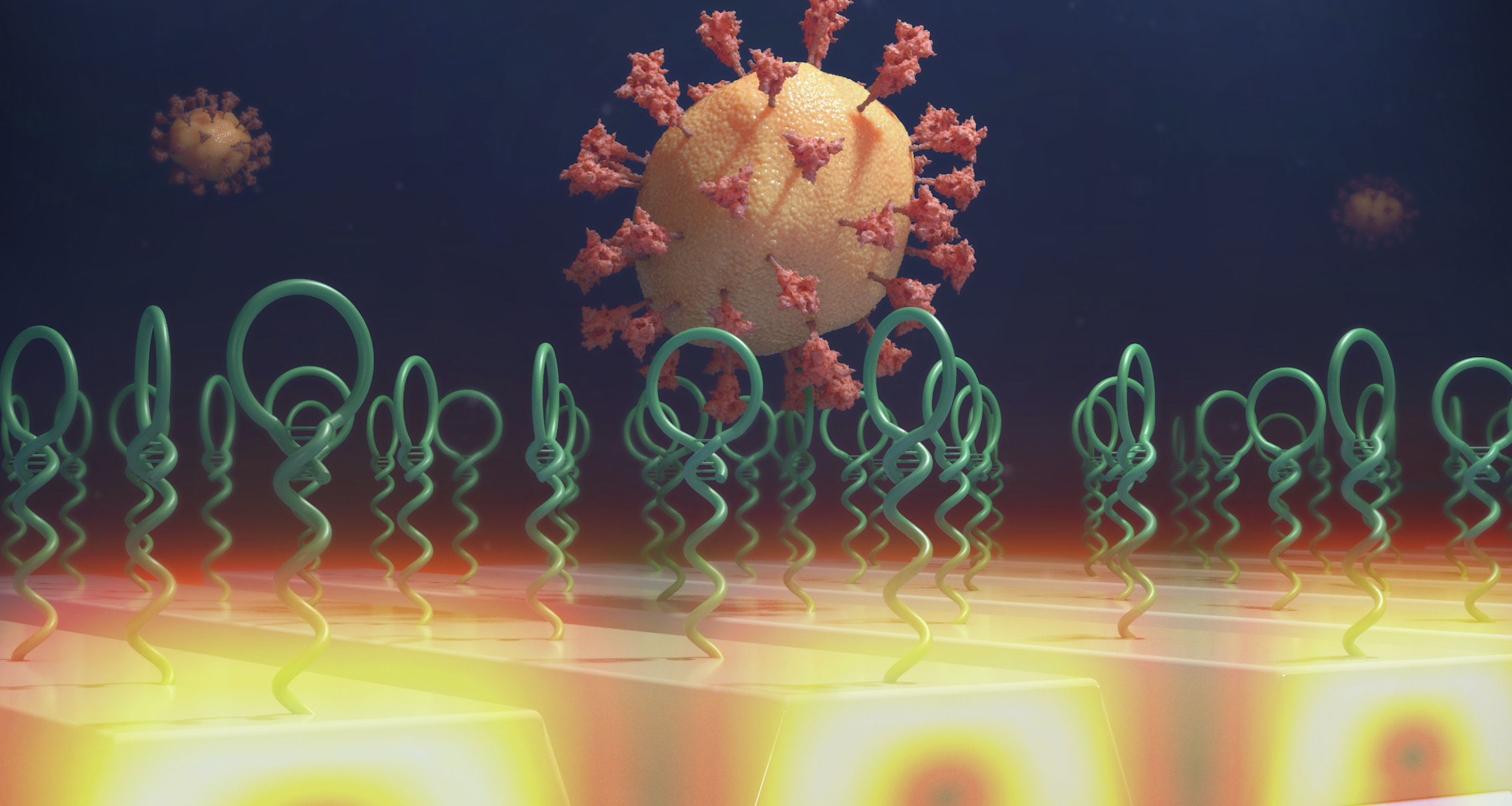
An artistic rendering of the NanoGripper’s applications. Sites on the gripper’s fingers recognize the spike protein of a virus, inset, and trigger fluorescent tags to emit light. When coupled with a sensor, individual viruses can be detected for a rapid COVID test, foreground. Alternately, the NanoGrippers can block viruses from entering cells by wrapping around the spike proteins, top right.

The NanoGripper could easily be engineered to target other viruses, such as influenza, HIV or hepatitis B, Wang said. In addition, Wang envisions using the NaoGripper for targeted drug delivery. For example, the fingers could be programmed to identify specific cancer markers, and grippers could carry cancer-fighting treatments directly to the target cells.

“This approach has bigger potential than the few examples we demonstrated in this work,” Wang said. “There are some adjustments we would have to make with the 3D structure, the stability and the targeting aptamers or nanobodies, but we’ve developed several techniques to do this in the lab. Of course it would require a lot of testing, but the potential applications for cancer treatment and the sensitivity achieved for diagnostic applications showcase the power of soft nanorobotics.”  

The National Institutes of Health and the National Science Foundation supported this work. Wang and Cunningham are affiliated with the Carl R. Woese Institute for Genomic Biology and the Holonyak Micro and Nanotechnology Lab at the U. of I. 

Editor’s note: To reach Xing Wang, email xingw@illinois.edu.  

The paper “Bioinspired designer DNA NanoGripper for virus sensing and potential inhibition” is available online.  DOI: 10.1126/scirobotics.adi2084

 This work was supported in part by NIH grants R21EB031310, R44DE030852 and R21AI166898. 

Two New Biosensing Instruments Take Big Leap Forward for Point-of-Care Diagnostics

Cancer Center at Illinois (CCIL) Program Leader Brian Cunningham has developed two new biomarker detection instruments with the potential to transform point-of-care diagnostics. In collaboration with researchers and clinicians at Stanford, Mayo Clinic, and Huntsman Cancer Institute, the Cunningham lab’s new instruments can vividly and rapidly detect a variety of cancer biomarkers using a novel gold nanoparticle tagging method.

“Nearly a decade ago, we invented a new biosensing instrument that incorporated our novel technology. We called it Photonic Resonator Absorption Microscopy (PRAM),” said Cunningham. “The original PRAM version we built was quite large, about four feet by eight feet, and cost approximately $200,000. It included an expensive microscope, spectrometer, cell incubator, and many top-of-the-line components. That first instrument did a great job of demonstrating the principle of PRAM, but the cost and size were prohibitive for clinical application. So, we’ve been working diligently to improve this technology for more practical applications in the lab and clinic.”

Brian and Weinan

Cancer Center at Illinois (CCIL) Program Leader Brian Cunningham (left) and graduate student Weinan Liu present two new biosensing instruments for enhanced biomarker detection. The Cunningham lab’s new PRAM Mini makes dramatic improvements in cost, size, and user-friendliness compared to the original PRAM prototype developed a decade ago.

Cunningham lab’s AuNP-tagging PRAM technology is fundamentally different than any other biomarker detection method available. 

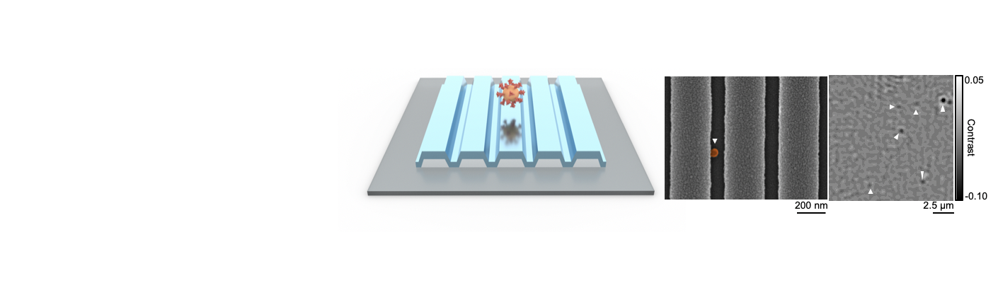
As opposed to a typical glass slide, the PRAM technique employs a photonic crystal as a surface for microscopy. The photonic crystal provides very high contrast for detecting and counting individual gold nanoparticles (AuNPs). The AuNPs are tags deposited on molecules for biosensing, enabling researchers to observe and count individual molecules in a sample. (See Figure 1 for explanation of nanoparticles.)

“In some cases, the molecule we may want to detect could be a protein, a nucleic acid molecule called micro-RNA, or a circulating tumor DNA. In other cases, we can detect antibodies, too,” said Cunningham. The potential for beneficial clinical application is far-reaching. Presently, the clinical researcher pioneering PRAM’s application is Cunningham’s collaborator Dr. Manish Koli at Huntsman Cancer Institute. Dr. Koli is driving the large genomic studies on prostate cancer patients, identifying specific biomarkers to differentiate between aggressive and non-aggressive cancer, and monitoring changes in biomarkers over the course of treatment.

Version 1: The Automated Portable PRAM (ap-PRAM)

Cunningham’s lab has been working diligently to reduce PRAM’s size, cost, and user-burden, and they are pleased to report two new PRAM instruments that accomplish this vision.

The first is an automated, portable version of PRAM named ap-PRAM. This novel instrument is reported in a recently published paper in the journal Biosensors and Bioelectronics.

PRAM is no longer bigger than a ping-pong table. The new ap-PRAM is roughly the size of a toaster oven with components costing about $12,000. Cunningham estimates it could be manufactured at scale with a cost of only $5,000. The ap-PRAM instruments’ advances include rapid biomarker detection, autofocusing, multiplexing, millimeter-scale tiled field of view, dynamic analysis of binding interactions, improved image processing algorithm, increased noise suppression, and a simplified representation of AuNPs’ binding and binary appearance in the enhanced digital resolution.

The ap-PRAM user doesn’t have to fiddle with knobs or manual adjustments, thanks to the microscope’s autofocusing which provides sharp display of AuNPs. The ap-PRAM can move the biosensor, too, by tiling together many fields of view and obtaining a larger surface area where all the AuNPs can be counted. It is also able to perform multiplexing—accommodating and measuring many biosensors in a single cartridge in rapid succession. Notably, ap-PRAM can also train its attention to one small point, measuring AuNPs accumulating there as a function of time. By studying the accumulation rate, this allows the user to ascertain the minimum time needed for accurate detection, rather than waiting for a full measurement of molecules.

“If the required information can be gathered in just five minutes, this time saved will greatly improve the point-of-care experience for both patient and user,” added Cunningham.

What is a nanoparticle

Version 2: The PRAM Mini

Because the cost and size of ap-PRAM would still be prohibitive for certain users, Cunningham’s lab wanted to push the boundaries even further. With significant efforts by graduate student researchers Kodchakorn Khemtonglang and Weinan Liu, the research team developed PRAM’s next iteration, the PRAM Mini. The new technology is reported in a recently published paper in the journal Biomedical Optics Express.

The PRAM Mini increases user-friendliness and dramatically reduces the form factor and costs of the original PRAM. Cunningham reports the PRAM Mini is the size of a textbook with an estimated manufacturing cost of only $500.

The PRAM Mini prototype is built on laser cut, quarter inch thick acrylic sheets, with holes in which are mounted a 3D printed structure to hold the optical components. The PRAM Mini eliminates the additional automated features of the ap-PRAM but is still capable of same limits of biomarker detection in other PRAM’s, using the team’s sophisticated AuNP tagging method. The PRAM Mini employs the same optical components as ap-PRAM but uses a Raspberry Pi microcomputer controller to communicates via Bluetooth with a linked mobile device.

“The PRAM Mini is very user-friendly, could easily go into a doctor’s office, and accomplishes detection within just five minutes. We believe it will revolutionize the future of point-of-care diagnostics for a range of different diseases,” Liu said.

While the original PRAM instrument provided high-contrast images, the more portable, user-friendly versions do come a sacrifice in image quality. However, with the incorporation of sophisticated machine learning algorithms, the research team is refining the technology so that affordability and portability don’t translate to a reduction in detection resolution.

A screen capture of PRAM’s digital biosensing. Anywhere there’s a AuNP, it absorbs red light and PRAM displays a little dark spot in the image. Instead of seeing many millions of AuNPs in aggregate, PRAM can count the individual molecules for unprecedented, accurate biomarker measurement. 

The Future of PRAM

In addition to continue to fine-tune the computational algorithms for maximal image resolution, the research team is considering additional improvements to the newest iterations of PRAM. “Because we want to keep the size and cost as minimal as possible without sacrificing ultimate biomarker detection efficacy, our next endeavor will be to work on the inclusion of multiple biomarker detections within a single image field of view—that is, maximizing information density given our existing constraints,” commented Cunningham.

In the future, when sitting in the doctor’s office, your care provider might use the non-invasive PRAM technology to perform accurate, cost-reducing cancer diagnostics within just five minutes. Your doctor might determine whether prostate cancer, for example, is an aggressive or less aggressive form. Or, your physician may use PRAM for treatment monitoring, assessing the efficacy of your treatment levels and deciphering any possible mutation recurrences that slipped by previous treatments.

Cunningham’s Product Development Ventures

To help distribute this new biosensing instrumentation at a larger scale, Cunningham formed a new company, Atzeyo Biosensors, with the aim “to change the lives of cancer patients” by developing innovative point-of-care diagnostic and monitoring platforms. Their innovative point-of-care technology will enable physicians to receive critical diagnostic results in real-time, in the office. Founded in 2023, Atzeyo recently completed an initial $300,000 round of seed funding to develop PRAM Mini into a commercial product. The company has organized a team of experts in diagnostics and detection instrument manufacturing, finance, and regulatory measures. Atzeyo will soon hire a product development company to take the initial PRAM Mini concept and design for manufacturability and user interface.

Summary

Cunningham’s innovative ap-PRAM and PRAM Mini instruments are fundamentally different than any existing biosensing technology for biomarker detection. PRAM employs a simple, low-input biochemistry method to label each molecule in the sample with a gold nanoparticle, enabling quick, accurate results. This technology holds great promise for cancer diagnostics and monitoring, as it can detect various cancer biomarkers, including proteins, microRNAs, and circulating tumor DNA. Finally, PRAM technology has the potential to transform point-of-care settings because of its low-cost instrumentation, simply assay method, and the high sensitivity obtained through the unique digital counting method.

“There’s nothing else that does what we are trying, so we think we have something special,” concluded Cunningham.

Editor’s notes:

The ap-PRAM is reported in the paper “Dynamic and large field of view photonic resonator absorption microscopy for ultrasensitive digital resolution detection of nucleic acid and protein biomarkers” and is available online.

doi.org/10.1016/j.bios.2024.116643

The PRAM Mini is reported in the paper “Portable, smartphone-linked, and miniaturized photonic resonator absorption microscope (PRAM Mini) for point-of-care diagnostics” ad is available online.

doi.org/10.1364/BOE.531388

Brian Cunningham is CCIL Program Leader for the Cancer Technology and Data Science (CTD) program, the Intel Alumni Endowed Chair of the Department of Electrical and Computer Engineering, a professor of bioengineering, and the Holonyak Micro and Nanotechnology Lab. Cunningham is also the Director of the Center for Genomic Diagnostics theme at the Carl R. Woese Institute for Genomic Biology.

To contact Brian Cunningham, email bcunning@illinois.edu

This story was written by Jonathan King, CCIL Communications Specialist.

Center for Genomic Diagnostics Receives First USDA Grant

Brian Cunningham, Intel Alumni Endowed Chair of Electrical and Computer Engineering, and Ying Fang, professor of pathobiology in the College of Veterinary Medicine (center) and lab members to develop portable point-of-use biosensor for detection of African swine fever virus in farm environments.

Brian Cunningham, Intel Alumni Endowed Chair of Electrical and Computer Engineering, and Ying Fang, professor of pathobiology in the College of Veterinary Medicine (center) and lab members to develop portable point-of-use biosensor for detection of African swine fever virus in farm environments. / Isaac Mitchell 

Foreign animal diseases are a global threat to swine production with the potential for detrimental economic implications. Recently, researchers at the University of Illinois Urbana-Champaign received a three-year grant of $650,000 from the U.S. Department of Agriculture to develop sensitive, rapid, low-cost, and portable point-of-use biosensors to improve on-farm detection and surveillance of African swine fever virus.

ASFV is a large DNA virus that infects swine and can result in a lethal hemorrhagic fever, spread rapidly to neighboring pigs, and cause excessive morbidity and mortality in swine populations. There currently is no effective vaccine or treatment for ASFV to help prevent infection and transmission. Further, detection of the virus is challenging because it relies on expensive offsite laboratory-based methods which often take too long for successful disease mitigation.

“ASFV is very important right now because it is already a deadly disease in other countries, and it can kill pigs quickly, usually within 7 to 10 days,” said Ying Fang (CGD/MMG), a professor of pathobiology in the College of Veterinary Medicine. “For field surveillance, if we have a portable device, we can take it to the field, and quickly detect ASFV-infected pigs. In this way, we can immediately apply the control and prevention measures.” 

With her expertise in animal diseases, Fang teamed up with Brian Cunningham (CGD leader), the Intel Alumni Endowed Chair of Electrical and Computer Engineering, to develop a biosensor for ASFV. Cunningham’s research focuses on developing nanotechnology-based biosensors for cancer and infectious diseases.

“We have been working on these technology approaches for about ten years, continuously refining and improving the biosensors, but mainly for cancer and infectious human diseases. So, when this USDA grant funding opportunity came up, Professor Fang encouraged us to try for it,” Cunningham said.

The project funding began on September 1 of this year, with the grant support coming from the USDA National Institute of Food and Agriculture’s Nanotechnology for Agriculture and Food Systems program. Over the next three years, the team will work on using genomics and proteomics to determine the diagnostic targets, specific viral nucleic acid sequences and proteins, for ASFV detection. Then, Fang’s research group will use their expert knowledge to develop and test laboratory-based methods for ASFV detection using this target. These methods will then be incorporated into Cunningham’s portable cartridge devices that use novel physics principles and nanotechnology methods to detect the target molecules from the virus. 

The work done at Illinois will focus on gene and protein level detection because research with live ASFV requires specialized facilities to eliminate exposure and transmission. To test their novel biosensor with active ASFV, Fang and Cunningham will collaborate with Jishu Shi, a professor of vaccine immunology at Kansas State University which houses the necessary biosafety level 3 facilities.

Overall, this grant represents a new portfolio of research for the Center for Genomic Diagnostics theme at the Carl R. Woese Institute for Genomic Biology. “We have been focused exclusively on human health and diseases and the underlying engineering science for sensing them. This represents how we have really strong pathobiology and veterinary medicine, combined with innovative engineering, here at Illinois. I think alone, neither of us would be able to do this project, but together, we make an excellent team,” Cunningham said.

“I hope this collaboration sends signals out university-wide that veterinary medicine is also important and an area that needs to be emphasized. So, I’m hoping to have more of this kind of collaboration and to continue developing new technologies to apply to livestock animals and veterinary medicine,” Fang said.

BY KATIE BRADY

September 30, 2024

https://www.igb.illinois.edu/article/center-genomic-diagnostics-receives-first-usda-grant

New photonic crystal approach can enable sensitive and affordable detection of biomarkers

Biomarkers are small molecules of interest to researchers, because they can indicate underlying diseases, often even before symptoms even appear. However, detecting these markers can be challenging as they are often present in very low quantities, especially in the early stages of a disease. Traditional detection methods, while effective, usually require expensive components like prisms, metal films, or optical objectives.

In a recent paper published in Applied Physics Letters, researchers at the University of Illinois Urbana-Champaign have unveiled a novel approach to detecting low concentrations of biomarkers that paves the way for biodetection technology that is simple to use, highly sensitive, and surprisingly affordable.

“The goal of this technology is early diagnostics, to be able to detect molecules associated with diseases at very low concentrations, sometimes a few molecules per millions, very early on,” said Seemesh Bhaskar, a postdoctoral researcher in the Cunningham lab and first author on the study. “Looking for very small concentrations of micro-RNA, circulating tumor DNA, and exosomes, for example, can help determine whether a patient will develop cancer one or two years down the line.”

Early detection of biomarkers is crucial for predicting and managing diseases effectively. There are many strategies for measuring the presence and concentration of biomarkers, but a common approach involves binding them with a fluorescent molecule, called a fluorophore, which emits fluorescence when excited with light.

Bhaskar noted that while there are technologies adept at detecting these low levels of fluorescent biomarkers, they are often bulky and expensive, limiting their accessibility in healthcare, particularly in resource-limited areas.

The approach encompasses a novel phenomenon for detecting light, called radiating guided mode resonance, which utilizes photonic crystals — thin pieces of glass with small gratings on the surface. These gratings help direct the photons, which are small particles of light, emitted from biomarkers along a pathway via a steering effect. This pathway is “tuned” to match the wavelength of the fluorescence emitted by the biomarkers, optimizing light collection and enhancing detection sensitivity.

Bhaskar likens this to a rhythmic dance of light energy within the crystal, where light is amplified while taking on the properties of the photonic crystal. One property of the crystal, called polarization selection, equalizes the polarization of the light, making for clearer and sharper detection of fluorescence. Together, this can result in an output that is 100 times stronger.

“To me, this is a whole new way of looking into the properties of light itself,” said Bhaskar. “The photons adapt, change, and evolve as they pass through the photonic crystal. The light picks up new characteristics without losing its essence. It’s a testament to the adaptability and transformative power of light.”

Discovery of this new phenomenon sets the stage for future detection platforms that will be able to detect molecules at picomolar levels without relying on costly components, making biodetection technology more sensitive, accessible, and affordable.

While radiating guided mode resonance can theoretically be used to enhance detection of many different biomarkers, the Cunningham lab is particularly interested in early cancer detection. The new phenomenon holds promise for affordable technology that will be critical for populations in resource-limited settings, where early disease detection and treatment can make a profound difference.

One of the lab’s long-term goals after development of this technology is to make it compatible with smartphones, further enhancing its accessibility. The envisioned future product would be a simple fixture attached to a smartphone’s camera, allowing a photonic crystal to illuminate a test sample while the phone’s camera measures the fluorescence emission.

“We are creating biosensing systems that are extremely sensitive while utilizing simple and inexpensive detection instruments,” said Brian Cunningham (CGD leader/MMG), a professor of electrical and computer engineering and program leader at the Cancer Center at Illinois. “This is what creates a path toward sophisticated health diagnostics making their way to our health clinics, farms, and homes.”

The study was funded by NIH, NSF, and the Cancer Center at Illinois. The paper can be found at https://doi.org/10.1063/5.0203999

Nanosensors Group
Holonyak Micro and Nanotechnology Laboratory
208 N. Wright St., MC-249
Urbana, IL 61801
(217) 265-6291
Log In