As COVID-19 continues to spread, bottlenecks in supplies and laboratory personnel have led to long waiting times for results in some areas. In a new study, University of Illinois, Urbana-Champaign researchers have demonstrated a prototype of a rapid COVID-19 molecular test and a simple-to-use, portable instrument for reading the results with a smartphone in 30 minutes, which could enable point-of-care diagnosis without needing to send samples to a lab.
The paper can be found on the Nanosensors Group “Publications” page, and at the Proceedings of the National Academy of Sciences website:
Researchers in Prof. Brian Cunningham’s Nanosensors Group at the University of Illinois, in collaboration with Prof. Singamaneni’s research group at Washington University, described a new approach for efficiently catalyzing chemical reactions using light, in the journal ACS Photonics. The researchers harnessed a new approach for amplifying electromagnetic fields in nanometer-scale volumes by coupling the energy from a laser into a nanostructured photonic crystal surface. The electromagnetic fields in the photonic crystal resonate with the laser’s wavelength, and when a metal nanoparticle is placed onto the photonic crystal, the electrons in the metal resonate as well. A portion of the resonating electrons become more reactive than ordinary electrons, and are able to transfer to nearby chemical molecules, and thus catalyze specific chemical reactions. The reactive electrons are often referred to as being “hot,” even though they are not hot in the temperature sense. The research shows that, by coupling laser light to a photonic crystal, chemical reactions are driven forward with greater efficiency, allowing less energy to perform a process than possible without the photonic crystal. Because the approach uses low illumination power that can be distributed over large surface areas, we envision the potential for optically driven chemical reactors. The reactors would be only several micrometers in height, with transparent windows, an inlet for precursors, an outlet for products, and nanoparticle-coated photonic crystals comprising the upper and lower surfaces.
ECE graduate student Qinglan Huang and IGB Fellow Taylor Canady led the research, which was performed in the Holonyak Micro and Nanotechnology Laboratory. The paper, entitled “Enhanced Plasmonic Photocatalysis through Synergistic Plasmonic–Photonic Hybridization“, by Q. Huang, T.D. Canady, G. Gupta, N. Li, S. Singamaneni and B.T. Cunningham, can be found at: ACS Photonics (2020).
The Nanosensors Group from the University of Illinois at Urbana-Champaign published a Critical Review for the journal Lab on a Chip, entitled “Digital Resolution Biomolecular Sensing for Diagnostics and Life Science Research.” The co-authors of the paper are Qinglan Huang, Nantao Li, Hanyuan Zhang, Congyu Che, Fu Sun, Yanyu Zhang, Taylor Canady, and Brian Cunningham. The paper reviews an important and very challenging frontier of the field of biosensing, where novel technologies are demonstrating the ability to digitally count molecules with single-unit precision. Through the use of enzymatic chemical reactions, nanoparticle tags, ultra-sensitive transducers, multiplexing detection instruments, and novel biochemistry, the reviewed technologies are finding applications in life science research, drug discovery, and diagnostics.
The paper can be found on the Nanosensors Group “Publications” page, and at the Lab on a Chip web site:
CHAMPAIGN, Ill. — Most viral test kits rely on labor- and time-intensive laboratory preparation and analysis techniques; for example, tests for the novel coronavirus can take days to detect the virus from nasal swabs. Now, researchers have demonstrated an inexpensive yet sensitive smartphone-based testing device for viral and bacterial pathogens that takes about 30 minutes to complete. The roughly $50 smartphone accessory could reduce the pressure on testing laboratories during a pandemic such as COVID-19.
“The challenges associated with rapid pathogen testing contribute to a lot of uncertainty regarding which individuals are quarantined and a whole host of other health and economic issues,” Cunningham said.
The study began with the goal of detecting a panel of viral and bacterial pathogens in horses, including those that cause severe respiratory illnesses similar to those presented in COVID-19, the researchers said.
“Horse pathogens can lead to devastating diseases in animal populations, of course, but one reason we work with them has to do with safety. The horse pathogens in our study are harmless to humans,” Cunningham said.
A schematic drawing of the new device system.
Graphic courtesy Brian Cunningham
The new testing device is comprised of a small cartridge containing testing reagents and a port to insert a nasal extract or blood sample, the researchers said. The whole unit clips to a smartphone.
Inside the cartridge, the reagents break open a pathogen’s outer shell to gain access to its RNA. A primer molecule then amplifies the genetic material into many millions of copies in about 10 or 15 minutes, the researchers said. A fluorescent dye stains the copies and glows green when illuminated by blue LED light, which is then detected by the smartphone’s camera.
“This test can be performed rapidly on passengers before getting on a flight, on people going to a theme park or before events like a conference or concert,” Cunningham said. “Cloud computing via a smartphone application could allow a negative test result to be registered with event organizers or as part of a boarding pass for a flight. Or, a person in quarantine could give themselves daily tests, register the results with a doctor, and then know when it’s safe to come out and rejoin society.”
There are a few preparatory steps currently performed outside of the device, and the team is working on a cartridge that has all of the reagents needed to be a fully integrated system. Other researchers at the U. of I. are using the novel coronavirus genome to create a mobile test for COVID-19, and making an easily manufactured cartridge that Cunningham said would improve testing efforts.
Study co-authors with Cunningham and Bashir were Fu Sun, Anurup Ganguli and Matthew B. Wheeler, of the U. of I.; and Ryan Brisbin and David L. Hirschberg, of RAIN Incubator; Krithika Shanmugam, of the University of Washington; and veterinarian David M. Nash.
The National Science Foundation and the Center for Genomic Diagnostics at the U. of I. ’s Carl R. Woese Institute for Genomic Biology supported this research.
Bashir also is the dean of the Grainger College of Engineering at Illinois.
Cunningham serves as a consultant to and owns stock in Reliant Immune Diagnostics, the company that licensed the technology described in this news release.
Editor’s notes:
To reach Brian Cunningham, call 217- 265-6291; email bcunning@illinois.edu.
The paper “Smartphone-based multiplex 30-minute nucleic acid test of live virus from nasal swab extract” is available online and from the U. of I. News Bureau. DOI: 10.1039/D0LC00304B
U OF I RESEARCHERS DEVELOPING SMARTPHONE APP FOR CORONAVIRUS TESTING
BRIAN MOLINE, ILLINOIS NEWSROOM | 4/3/2020 3:30:07 PM
A team of University of Illinois researchers is working on a smartphone application that would detect the novel coronavirus within 30 minutes, without the need for a diagnostic lab.
Illinois Newsroom’s Brian Moline spoke with Brian Cunningham. He’s a professor of electrical and computer engineering, and a professor at Nick Holonyak Jr., Micro and Nanotechnology Lab on the Urbana campus.
He said that’s just one of several diagnostic technologies that his research team is working on in collaboration with others.
“One test we’re developing is based on detecting the nucleic acid sequence, part of the genome of the virus,” Cunningham said. “In that test, you take the virus, break it apart by putting it into a chemical that breaks the virus membrane and releases the RNA material. And then, in that test, you take that genomic sequence, you have a special molecule that recognizes it, and then turns each individual piece of the genome into thousands and then millions of individual copies that fluoresce. That test is performed inside a small, plastic cartridge that has an output that is readable by a phone.”
Cunningham said there are both advantages and disadvantages in working with smartphones.
“Of course, now, everybody has one,” he said. “It’s a powerful device that you can use in many ways, but you can also use it in measuring biological diagnostic tests. The downside is that it’s not approved by regulatory agencies to use a mobile device like that, for doing a test, and you have to show very rigorously that a phone-based system can give results that are equivalent to laboratory tests using clinical samples.”
Cunningham said his group has completed successful testing of the smartphone application using respiratory viruses from horses. He has submitted the research to the journal Lab on a Chip to be considered for fast-track publication. You can read that submission below.
Cunningham said they have also submitted applications for emergency funding to the National Science Foundation and the National Institutes of Health.
In addition to the smartphone application, Cunningham said his research team is also working on a biosensor method of testing that detects and counts the intact coronavirus from nasal swab extract. He said they are also working on another test that would detect if a person has antibodies that could give them immunity to COVID-19.
A new research center at the University of Illinois directed by Donald Biggar Willett Professor in Engineering Brian Cunningham aims to revolutionize diagnostics and personalized medicine, developing technologies that are at once more accurate, more affordable, and more practical for routine care.
The Carl R. Woese Institute for Genomic Biology (IGB) and the Grainger College of Engineering are working together to support the launch the new Center for Genomic Diagnostics (CGD), which will be housed within the IGB. The CGD will also take advantage of specialized laboratory space and equipment in Illinois’ Holonyak Micro and Nanotechnology Lab.
The vision for the new center begins with molecules called biomarkers that are naturally produced as part of a healthy biological state or disease process. If such a molecule is produced in detectably larger or smaller quantities in certain conditions, it can serve as the basis for a reliable test for that condition. For example, the hormone human chorionic gonadotropin, which is detected in urine by home tests during early pregnancy, is a biomarker. Other biomarkers signal the presence of diseases, including some types of cancer.
“Our goal for the center is first to use genomics and bioinformatics to identify novel biomarkers,” Cunningham said. “As we seek to validate how biomarker presence and concentration changes with a specific health condition, we’re also interested in developing novel biochemistry methods for selectively detecting those molecules with methods that are simple, yet extremely sensitive.”
The concept for the center emerged from Cunningham’s research theme at the IGB, Omics Nanotechnology for Cancer Precision Medicine, which was established in 2016. The theme brings together computational, biochemical, biomedical, and engineering approaches to biomarker discovery and innovations in the realm of biomarker detection. In its conversion from a theme to a center, members are broadening their focus on cancer to include a wide variety of diseases and conditions.
“There are biomarkers for diseases other than cancer, as well as biomarkers that provide information on a person’s nutrition, environment, microbiome, and metabolism,” Cunningham said. “There are genomic biomarkers for infection and immunity, for inflammation and sepsis, and surprisingly even for environmental exposure, psychological stress, and anxiety . . . So that’s how we’re turning from a cancer group into a genomic diagnostic center, so we can consider things more broadly.”
The center already works closely with the Cancer Center at Illinois. With its expanded scope, the CGD is also growing its relationships with Illinois’ Health Care Engineering Systems Center, as well as the Mayo Clinic and Illinois Alliance.
“Our diagnostic [capability] is one of the things that allows medicine to be personalized,” Cunningham said. “So if you can have a test for a biomarker that tells the clinician something about a particular patient, they have a specific gene is being expressed, or they have a protein molecule that is present in high levels, the information can indicate that the patient is more likely to have successful treatment with a particular drug.”
The faculty and research staff that will join the center from the original IGB research theme and from around campus represent a diversity of backgrounds and expertise.
“Our team includes faculty with backgrounds that span bioinformatics, biochemistry, chemistry, biology, and nutrition, along with engineers like myself whose specialty is detection instrumentation and biosensors.” Cunningham said; the center also involves clinicians from Mayo Clinic, Carle, OSF Hospital, Stanford, and Huntsman Cancer Institute “who could give us guidance about what kind of information would better guide their treatment decisions. Our clinical partners also inform us about the shortcomings of currently available technologies—and help us target our work to where it can have the greatest clinical impact.”
The center was initiated in January 2020, and is planning a symposium to showcase its research goals later this spring.
CHAMPAIGN, Ill. — A fast, inexpensive yet sensitive technique to detect cancer markers is bringing researchers closer to a “liquid biopsy – a test using a small sample of blood or serum to detect cancer, rather than the invasive tissue sampling routinely used for diagnosis.
Researchers at the University of Illinois developed a method to capture and count cancer-associated microRNAs, or tiny bits of messenger molecules that are exuded from cells and can be detected in blood or serum, with single-molecule resolution. The team published its results in the Proceedings of the National Academy of Science.
“There are specific microRNA molecules whose presence and concentration is known to be related to the presence and aggressiveness of specific types of cancer, so they are known as biomarkers that can be the target molecule for a diagnostic test,” he said.
Cunningham’s group developed a technique named Photonic Resonator Absorption Microscopy to capture and count microRNA biomarkers. In collaboration with professor Manish Kohli at the Moffitt Cancer Center in Florida, they tested PRAM on two microRNAs that are known markers for prostate cancer.
They found it was sensitive enough to detect small amounts that would be present in a patient’s serum, yet also selective enough to detect the marker among a cocktail of molecules that also would be present in serum.
“One of the main challenges of biosensing is to maintain sensitivity and selectivity at the same time,” said Nantao Li, a graduate student and co-first author. “You want it to be sensitive enough to detect very small amounts, but you don’t want it to pick up every RNA in the blood. You want this specific sequence to be your target.”
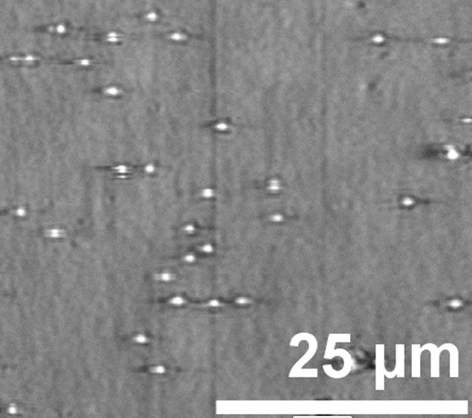
Each dot seen in this PRAM image represents one microRNA that has bound to the sensor.
Image courtesy of Nantao Li
PRAM achieves both qualities by combining a molecular probe and a photonic crystal sensor. The probe very specifically pairs to a designated microRNA and has a protective cap that comes off when it finds and binds to the target biomarker. The exposed end of the probe can then bind to the sensor, producing a signal visible through a microscope.
Each individual probe that binds sends a separate signal that the researchers can count. This means researchers are able to detect much smaller amounts than traditional methods like fluorescence, which need to exceed a certain threshold to emit a measurable signal. Being able to count each biomarker also carries the added benefit of allowing researchers to monitor changes in the concentration of the biomarker over time.
“With PRAM, we squirt a sample into a solution and get a readout within two hours,” said postdoctoral researcher Taylor Canady, a co-first author of the study. “Other technologies that produce single-molecule readouts require extra processing and additional steps, and they require a day or more of waiting. PRAM seems like something that could be much more feasible clinically. In addition, by using an optical signal instead of fluorescence, we could one day build a miniaturized device that doesn’t need a trained laboratory technician.”
The PRAM approach could be adapted to different microRNAs or other biomarkers, the researchers say, and is compatible with existing microscope platforms.
“This approach makes the idea of performing a ‘liquid biopsy’ for low-concentration cancer-related molecules a step closer to reality,” Cunningham said. “This advance demonstrates that it is possible to have an inexpensive and routine method that is sensitive enough to require only a droplet of blood. The results of the test might tell a physician whether a regimen of chemotherapy is working, whether a person’s cancer is developing a new mutation that would make it resistant to a drug, or whether a person who had been previously treated for cancer might be having a remission.”
The Carl R. Woese Institute for Genomic Biology at the U. of I. and the National Institutes of Health supported this work. Illinois chemistry professor Yi Lu and bioengineering professor Andrew Smith were coauthors of the work.
Editor’s note: To reach Brian Cunningham, call 217-265-6291; email bcunning@illinois.edu. The paper “Digital-resolution detection of microRNA with single-base selectivity by photonic resonator absorption microscopy” is available online. DOI: 10.1073/pnas.1904770116
Infectious diseases such as Zika and Dengue remain a top contributor to death and disability across the globe, according to the World Health Organization. Diagnosing and treating these diseases, which often have similar symptoms, is especially difficult in developing countries, where access to health care and laboratories is often limited.
Mosquitoes can carry a variety of infectious diseases that until now were difficult and expensive to diagnose.
Researchers at the University of Illinois at Urbana-Champaign are developing a lab-on-a-smartphone system that will enable healthcare professionals to detect disease at the point of care. Funded by the National Institutes of Health, the 4-year grant will enable the team to develop a handheld instrument that can detect and report the presence of pathogens in less than 30 minutes using a single drop of blood – all with a smartphone clip-on instrument that costs less than $10.
“This device can substantially reduce the time, cost, and inconvenience of doing a standard lab test, while still incorporating all the controls that make the test valid,” said Principal Investigator Brian Cunningham, Director of the Holonyak Micro & Nanotechnology Lab and a Donald Biggar Willett Professor in Engineering at Illinois. “The information can then be shared immediately with an online health care provider who can make decisions about treatment.”
The system will comprise three components: a microfluidic cartridge, a clip-on instrument, and a smartphone with a rear-facing camera. The phone’s software will be able to connect with a cloud-based service system that detects and reports the presence and concentration of a panel of viral pathogens.
The microfluidic cartridge will contain pre-dried “primers”, or short nucleic acid sequences, that specifically recognize and amplify
Brian Cunningham
DNA from the pathogen targets. When the clip-on instrument interfaces with the cartridge – similar to putting a credit card into a credit card reader – the instrument will use LED illumination to excite fluorescent dyes within the cartridge. The camera will capture a movie of the reaction while it takes place, utilizing algorithms that can detect, track, and count the individual copies of DNA that are specific to each virus. The phone will then link to a cloud-based system to interpret the results and forward them to an offsite medical professional.The project involves numerous technical challenges, one of which is creating a device that provides a reliable result even with a very small amount of blood. Another challenge is developing robust and accurate tracking algorithms for this new type of video data.
“We will have to model the signal and noise, due to many non-ideal conditions in the field, to come up with an optimal processing algorithm,” said Minh Do, a co-investigator and professor of electrical and computer engineering at Illinois, who is leading the image and video processing work. The research could result in other smartphone biosensing applications, he says.
Other research contributors include Grainger College of Engineering Dean Rashid Bashir, a faculty member in Illinois’ Bioengineering Department, and Ian Brooks, a research scientist in Illinois’ School of Information Sciences.
Once the group has a working prototype, they plan to take the device to a clinic in Brazil to test it in a real-world environment.
“Even in the United States, where we enjoy good access to diagnostic testing laboratories, it can take hours or days to have results from lab work,” Cunningham said. “We believe this can provide affordable access and fast results to patients around the globe.”
Original story posted on the Holonyak Micro and Nanotechnology Laboratory site:
Harnessing light’s energy into nanoscale volumes requires novel engineering approaches to overcome a fundamental barrier known as the “diffraction limit.” However, researchers in the Cunningham Group have breached this barrier by developing nanoantennas that pack the energy captured from light sources, such as LEDs, into particles with nanometer-scale diameters, making it possible to detect individual biomolecules, catalyze chemical reactions, and generate photons with desirable properties for quantum computing.
See the full press release at:
https://ece.illinois.edu/newsroom/article/34397
For full details, see our publication in Nano Letters:
“Microcavity-Mediated Spectrally Tunable Amplification of Absorption in Plasmonic Nanoantennas,” Q. Huang and B.T. Cunningham, Nano Letters, in print 10.1021/acs.nanolett.9b01764.
A three-time Illinois alumnus (BSEE 1950, MSEE 1951, PhD 1954), Holonyak’s GaAsP red alloy LED paved the way for today’s solid-state lighting revolution that is replacing Edison’s incandescent bulb and other less efficient light sources.
After productive stints with Bell Labs and GE and service in the U.S. Army Signal Corps in Japan, Holonyak joined the Illinois faculty in 1963, establishing a research program in the Electrical Engineering Research Lab.
For the next forty years, Holonyak and his students would continue to produce major technology advancements such as the world’s first quantum-well laser, the impurity-induced layer disordering technique for high-power laser, and the stable native oxide for vertical-cavity surface-emitting lasers (VSCELS).
These advancements led to brighter and more efficient LEDS and lasers which are used in modern fiber-optic communications, CD and DVD players, optical storage, medical diagnosis, surgery, ophthalmology, and other applications.
A photo gallery from the ceremony can be found here.